Light sheet microscopy has become very popular for imaging living cells and organisms due to its imaging speed and low photo-toxicity. There are a large number of microscopes that are based on similar technology. So far, commercial solutions implement only some of these capabilities. Thanks to the OpenSPIM project, it is possible to build a light sheet microscope in the lab and select specific components that are ideal for imaging a particular sample. The control and acquisition of the instrument built according to this guide is done using the freely available MicroManager software, for which a special plugin has been created.
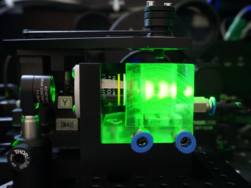
SPIM(Selective Plane Illumination Microscopy) microscopy is a non-invasive method whose principle consists in illuminating the sample with a laser beam stretched into a line(light sheet), using a cylindrical lens and a vertical aperture. The detector(camera) is positioned perpendicular to the beam and therefore a 2D image is projected onto it from the focal plane. Thanks to this arrangement, noise in the form of a signal originating outside the focal plane can be suppressed and an image approaching the quality of confocal microscope images can be obtained. In addition, very high scanning speeds can be achieved, with the only limitation being the scanning speed of the cameras.
By moving the sample, optical sections can be imaged and then a 3D reconstruction can be created. The sample can be rotated sequentially and images taken at different angles, which are then digitally reconstructed and stacked into the resulting "multi-view" 3D model. This contains high-resolution information about the entire sample. As this is a wide-field microscopy, light of very low intensity (mW/cm2) is incident on the sample, and only on a limited area. These features, together with the high scanning speed, make the SPIM microscope an ideal instrument for imaging living cells and organisms.
| Components | Product | Features |
|---|---|---|
| Laser | OBIS | Wide range of wavelengths |
| Camera | Zyla / iXon | High sensitivity and fast scanning |
| Incubation chamber |
Custom manufacturing |
Dimensions according to customer requirements |
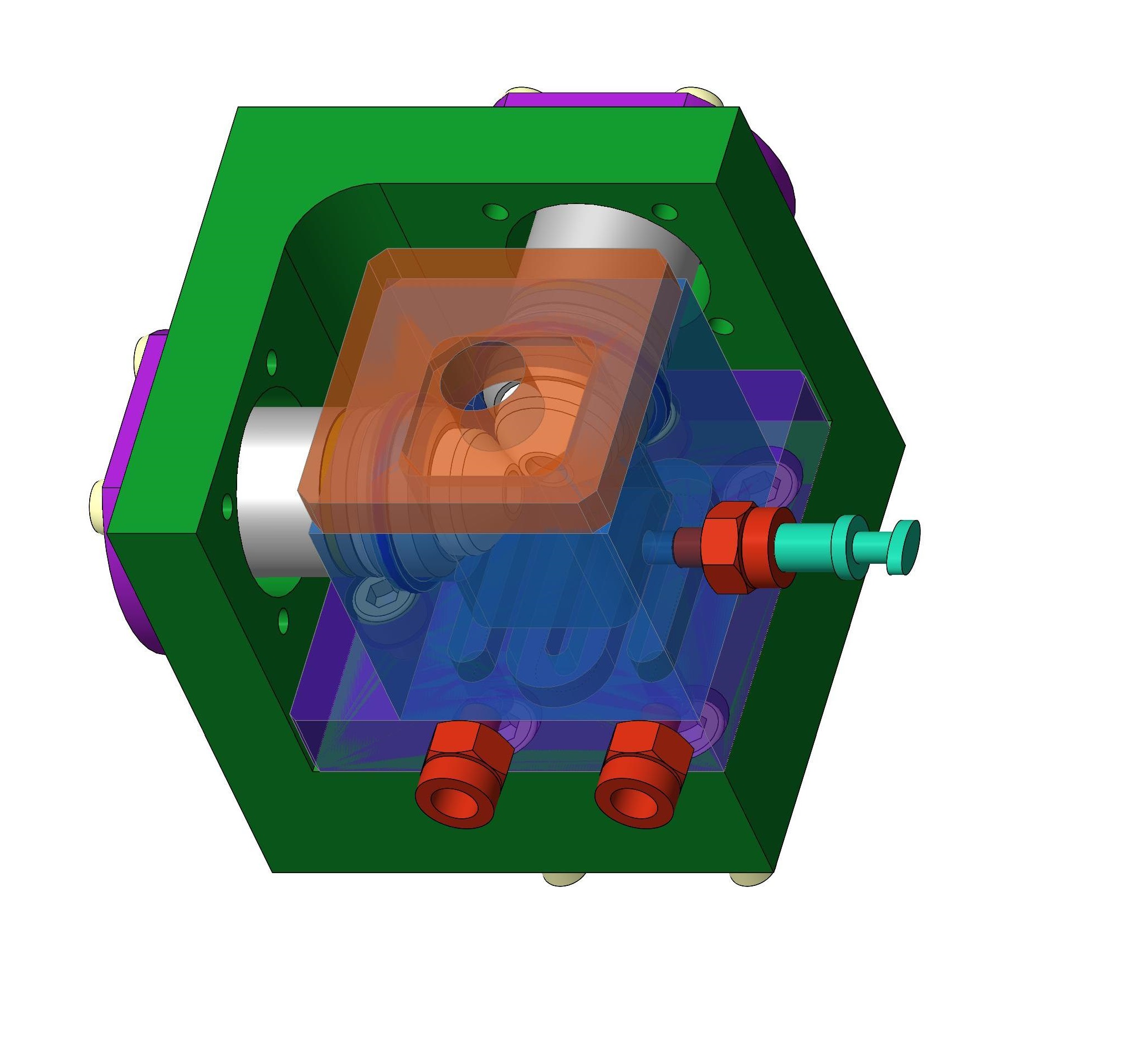
| Positioning unit |
4D Stage | Three-axis movement and rotation |
| Optics | Telescope, tube lenses | Effect of laser beam diameter and magnification on the emission path |
| Optical filters | Excitation filters and emission filters | Filtration of specific wavelengths |
| Anti-vibration pad | CSP | Vibration damping for longer experiments |
| Acquisition software | MicroManager | Microscope control |
Key benefits of the SPIM microscope:
- Low purchase price compared to commercially available systems
- High scanning speed fully dependent on camera characteristics
- Low level of toxicity of incident light on the sample
- Very flexible configuration depending on the requirements of the designer