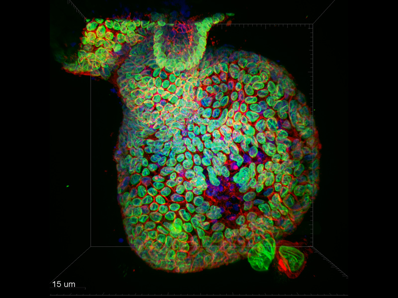
Figure 1: Organoid simulating mouse intestinal tissue. DNA (blue), LaminB1 (green, nuclear envelope), GM130 (red, cis-golgi). Confocal image taken on a Dragonfly microscope using an iXon Ultra 888 EM CCD camera - 20× immersion objective, 40 μm pinhole slit. Ronan Mellin, Dr Luke Boulter, MRC Human Genetics Unit
Spatial cell cultures of pluripotent stem cells enable the formation of "organoids". These are complex cellular structures that resemble organs and provide new opportunities for drug testing, assessment of tissue regeneration and observation of other in vivo biological processes (gene mutations, organ development and damage, cancer). Mouse and human stem cells have been used to generate many types of organoids that serve as models of intestinal epithelium, liver tissue and brain.
The potential use of organoids is demonstrated by a study in which cells were isolated from colorectal cancers (CRC) from real patients. The organoids that emerged from these CRC cells showed a high degree of similarity to the original CRCs in several characteristics, allowing direct testing of potential anti-cancer drugs. Organoids from different patients were multiplied and sorted into a so-called biobank, which serves as a resource for further analysis. For example, it is possible to determine the relationship between certain drugs and the genetic material of a particular organoid. Further development of this application will lead to methods for personalised treatment of different types of cancer. At the same time, due to their complexity, organoids will fill the gap between in vitro research of new drugs in 2D cell cultures and clinical studies.
One important method for analyzing these cellular structures is microscopy. Due to the diversity of organoids, it is essential that the microscope used provides sufficient flexibility. As this is live cellimaging, emphasis is also placed on reducing the toxic effects of light andbleaching. In addition, due to the size of these spatial cell cultures, high scanning speed and sufficient penetration depth are required (Andor, 2016).
The Dragonflyconfocal microscope meets all the requirements for imaging organoids:
- Flexibility of confocal imaging - zoom optics on the cameras, two pinhole sizes on the disk(SDCM)
- High frame rate - 103 frames per second (2048 × 2048 pixels) - Zyla 4.2 Plus sCMOS camera
- Low light intensities - extremely sensitive cameras (QE > 90%) - iXon Ultra 888 EM CCD camera
- "Multimodal" imaging - confocal microscopy, widefield microscopy, TIRF microscopy
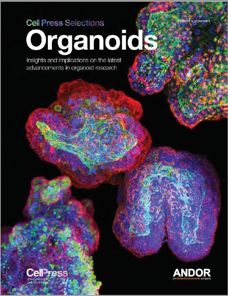
Cell Press Selection - Organoids
- A collection of the most important recent scientific articles on Organoids published in Cell
- Engineering Stem Cell Organoids
- Complex Tissue and Disease Modeling using hiPSCs
- Modeling Development and Disease with Organoids
- and many others
- To download, please fill out the short form that appears when you click on the image:
Source of the report, "Organoids in the Study of Liver Development and Cancer." Accessed November 25, 2016 www.andor.com