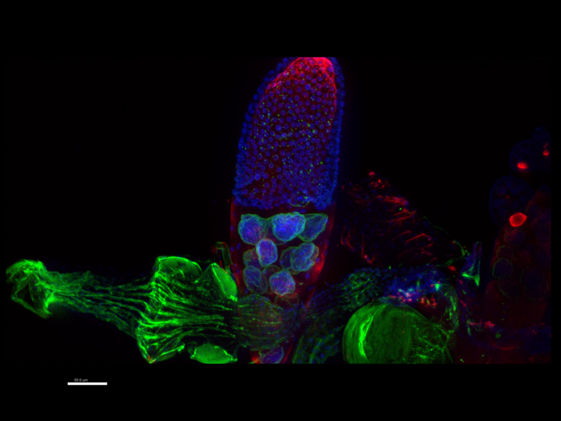
The DOT(Dresden Ovary Table, Tomancak lab/MPI-CBG Dresden) project maps RNA localization during oogenesis in Drosophila. The main method of this research project is the so-called FISH(Fluorescence in situ hybridization). This method allows the visualization of specific molecules that are complementary to the fluorophore used. In the case of the DOT project, these are mRNA molecules that provide information on gene expression at different developmental stages of the octomycete (Jambor et al., 2016).
The DOT database contains data from more than 8000 in situ hybridization experiments. A Revolution DSD2 confocal microscope from Andor was used to analyze and 3D visualize these experiments. The database contains 32,000 3D images, revealing the localization of more than 1700 genes in octopus ovaries at all stages of oogenesis. Studies requiring the acquisition of such a large number of 3D images can only be realised thanks to the high speed and high quality of imaging made possible by SDspinning disc confocal microscopy technology in conjunction with extremely fast sCMOS cameras (Andor Zyla).
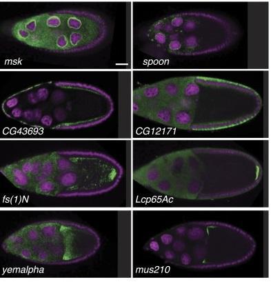
Figure 1: Sample gene expression patterns
The main advantages of the current Andor Revolution DSD2 confocal microscope:
- Immediate and fast switching between widefield and confocal microscopy
- High confocal scanning speed - 22 fps (928 × 2048 pixels)
- LED(Laser free) light source with 370-700 nm range
- Compatible with most microscopes, macroscopes and stereomicroscopes from the world's leading manufacturers
- Includes Andor Zyla's extremely sensitive and fast sCMOS cameras
News source: "Dresden Ovary Table." Accessed November 28, 2016 tomancak-srv1.mpi-cbg.de
News Source: Jambor, Helena, Vineeth Surendranath, Alex T Kalinka, Pavel Mejstrik, Stephan Saalfeld, and Pavel Tomancak. "Systematic Imaging Reveals Features and Changing Localization of mRNAs in Drosophila Development." eLife 4. Accessed November 28, 2016. doi:10.7554/eLife.05003


