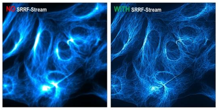
A new, very fast and flexible method for super-resolution microscopy that can be operated, due to its applicability over a very wide range of fluorophore densities and excitation strengths, even with conventional fluorophores(e.g. GFP) under low illumination intensities.
Depending on the parameters, a high resolution of 50-150 nm can be achieved with excitation strengths more than 106 times lower than typical for conventional localization microscopy methods. With a super-resolution scan rate of >10 frames per second and real-time output, the SRRF Stream can be used to study fast, dynamic physiological processes in living cells at the single molecule level.
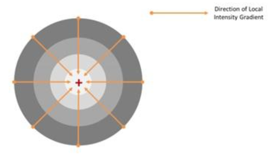
SRRF uses the concept of Radiality Arrays as a first important step to achieve super-resolution, where radial transitions at each point in the image are analyzed at an interpolated user-defined resolution that is greater than the resolution of the natural pixel of the receiving camera.
The result is a complex data region containing a large amount of information about local intensity gradients that provides detailed information about the exact location of fluorophores within each image. In the diagram, the fluorescence marker (red cross) is the center of the intensity gradient field, as the point of maximum radial symmetry. The SRRF process requires multiple images to be captured and composited simultaneously (typically 50-100) to derive a single higher resolution output image. The SRRF Stream method is primarily tied to the use of Andor's EMCCD cameras, specifically the iXon Life and iXon Ultra, which can be installed on any microscope without the need to acquire expensive systems designed for super-resolution microscopy.


