We distribute this product only in the Czech Republic and Slovakia.

RM5 confocal Raman microscope

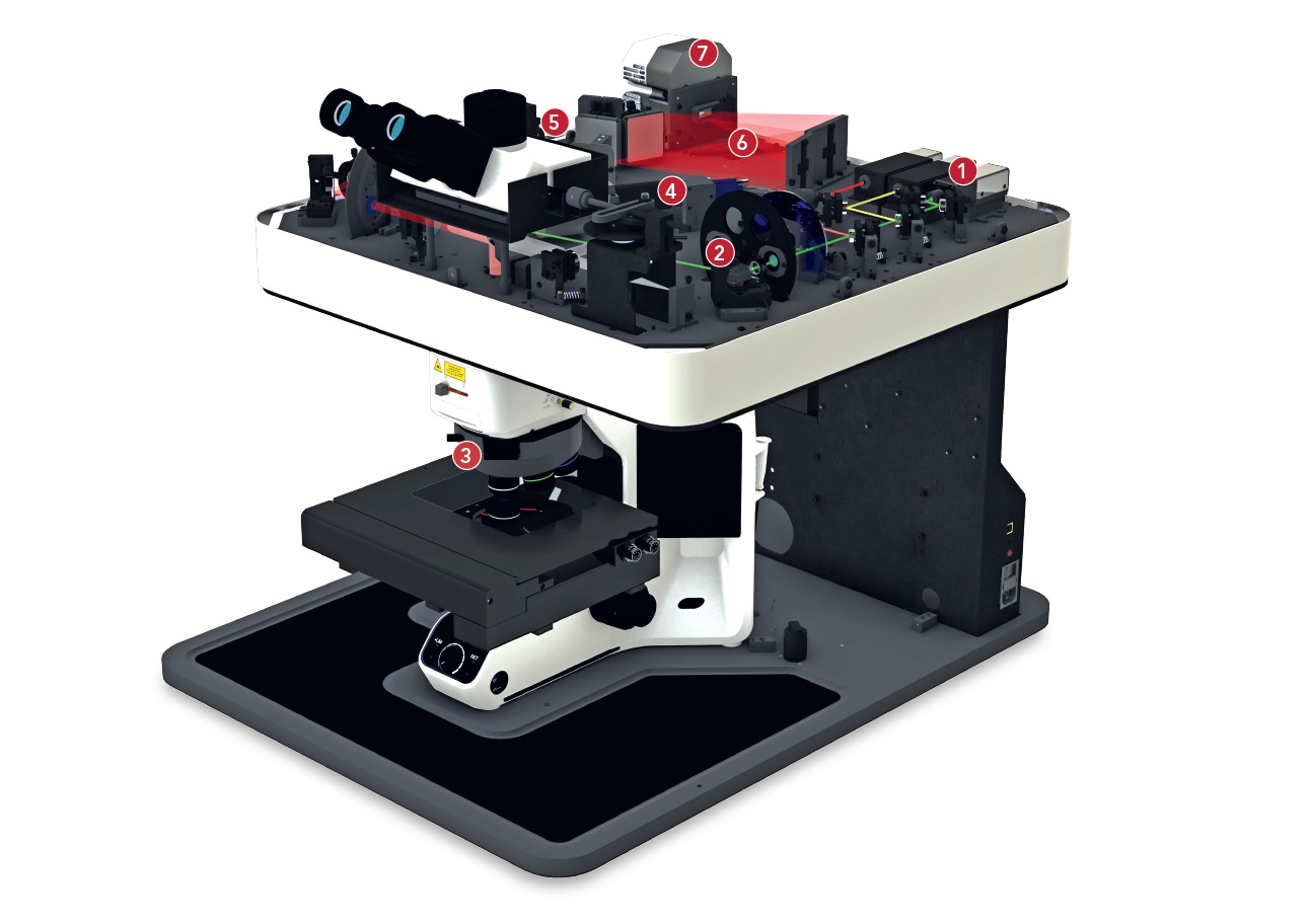
The new RM5 model from Edinburgh Instruments is a compact, fully automated confocal Raman microscope that excels in high precision spectral resolution (1.4 cm-1 FWHM) over the entire spectral range of 50 cm-1 - 4000 cm-1, high spatial resolution to resolve details down to 1 µm, and high measurement sensitivity to detect even the faintest Raman signals. Thanks to the full automation of the entire instrument, the desired Raman data can be obtained quickly, very simply and without the need for manual intervention and adjustment.

Key features:

  • Up to three computer-controlled lasers can be integrated - wavelengths from 400 nm to 1064 nm
  • Software selection of laserwavelengths and laser power settings
  • Confocality for high resolution and suppression of background signals caused by fluorescence
  • Software adjustable pinhole and slit size to provide confocality and high spatial resolution in all 3 dimensions
  • Ability to integrate up to 2 high efficiency and sensitive detectors - CCD, EMCCD or InGaAs
  • Exceptional data acquisition speed - up to 0.001 seconds for a complete spectrum due to the combination with EMCCD
  • Integrated spectrograph with asymmetric Czerny-Turner design and 225 mm focal length
  • 5 position diffraction grating carousel
  • Motorized carousel with 4 positions for three Raman filters and one beam splitter
  • Integrated internal standards and auto-calibration procedures (Neon and Si standard) - no need to add external sources
  • Automatic laser beam alignment and laser beam correction using piezoelectrically shifted mirrors
  • Olympus BX53 high-performance upright microscope providing all visualization and contrast techniques - brightfield, darkfield, polarized light, DIC and fluorescence
  • Fully motorized, software-controlled stage for sample mapping in X, Y and Z axes (XY manual stage also available)
  • Microscope stage heating/cooling capability
  • Raman mapping capability - analysis of chemical and physical properties across the sample, recording of sample changes over time, etc.
  • Built-in high-resolutionCMOS camera for live-sample imaging
  • Option to add a second CMOS camera for even higher resolution and stitching of Raman mapping images
  • User-friendly Ramacle® software for complete system control, data acquisition, display and analysis
  • Optional upgrade module for chemometric analysis and more
  • Option for polarization analysis of Raman spectra
  • System supplied with laser blocking when the instrument cover is opened - Class I safety class, or without laser cover - Class 3B safety class
EDINBURGH INSTRUMENTS - Logo
EDINBURGH INSTRUMENTS

Aplikační oblasti:

Biological Sciences | Pharmacy | Chemistry, Analysis and Control of Chemicals | Polymers | Nanomaterials | Cosmetics | Semiconductors | Art & Museum - Material Analysis | Forensic Analysis | Geology

Miroslav Kořínek
Expert advisor

RNDr. Miroslav Kořínek Ph.D.

+420 720 994 252

korinek@optixs.cz

Send inquiry

OptiXs care

  • We will expertly consult your planned application
  • Our team is able to integrate the product into a larger system
  • We ensure fast delivery of spare parts and local service
What else we can help with

Popis produktu

RM5 - princip

The excitation laser beam from one of the three integrated lasers (1) is guided directly onto a movable XYZ microscope stage ( 3) via a series of motorized mirrors with piezoelectric displacement, where it is focused onto the sample by an objective lens. The sample can be monitored in real time using a built-in CMOS camera (4). The same lens is then used to collect the stray light generated from the sample and a filter is used to remove unwanted stray light from the area outside the laser beam focusing area. The transmitted Raman scattered light passes through an adjustable confocal pinhole (5) before entering the spectrograph. The light is then split, by one of the 5 diffraction gratings located in the fully motorized carousel, into its fundamental wavelengths (6), which are focused into the detector/detectors (7) and displayed as a spectrum. All internal standards (2) regarding wavelength calibration, Raman shift, sensitivity and automated laser beam positioning are included in the Ramacle® control software.

Accessories

Lasers

The microscope includes upto three integrated narrowband lasers with wavelengths of 532 nm, 638 nm, 785 nm (but all wavelengths from 400 nm to 1064 nm are available), the selection and adjustment of the laser beam intensity is fully controlled in the software. The ability to select the appropriate excitation wavelength, optimise it for the application and the presence of a confocal pinhole ensures suppression of background signals that may be caused by fluorescence when analysing biological samples. Automated laser beam positioning (laser alignment) and wavelength calibration is one of the internal standards integrated into the RM5 microscope at the time of manufacture.

Optics

The main optical element of the RM5 microscope is a fully automated carousel with a total of 4 positions for three dichroic filters (edge and notch) and one beam splitter and an adjustable confocal pinhole. Filter exchange and selection, beam splitter position and pinhole size adjustment is enabled in the control software. Automatic alignment of the instrument is provided by two built-in piezoelectrically shifted mirrors. An optional accessory for advanced polarization analysis of Raman spectra is an additional polarizer and analyzer allowing the study of vibrational mode symmetry, single crystal orientation, polycrystalline samples and anisotropic materials.

Spectrograph

The system includes a spectrograph with a proven Czerny-Turner design and a focal length of 255 mm consisting of a continuously adjustable slit and a carousel on a diffraction grating with 5 positions to cover the widest possible spectral range (up to 4000 cm-1). All calibration standards are implemented directly into the control software during manufacture. These internal standards are mainly:

  • Wavelength calibration (Neon standard),
  • Raman shift calibration (Si standard),
  • Sensitivity verification (Si standard),
  • Automated laser beam positioning

Detectors

The RM5 microscope includes an integrated, high-sensitivity CCD detector for ultra-low noise, with thermoelectric cooling down to -60 °C and a resolution of 1650 x 200 pixels or 2000 x 256 pixels (for increased sensitivity and spectral range). A second port is available for integration of an additional detector with complementary spectral coverage - EMCCD or InGaAs - for increased speed, sensitivity and sampling rate. The RM5 ensures optimal performance through a combination of highly sensitive and fast detectors and provides exceptional data acquisition speed - a complete spectrum can be recorded in 0.001 seconds.

Evidence of maximum sensitivity is demonstrated by the detection of a fourth-order silicon band at 1940 cm-1 (see silicon spectrum after 785 nm laser excitation).

Spektrum křemíku po excitaci 785 nm laserem

High-performance microscope

The latest generation of theOlympus BX53 series upright microscope , which is part of the RM5 system, allows the use of all advanced visualization and contrast techniques for sample imaging, which include brightfield, darkfield, polarized light, Norman differential interference contrast (DIC) and fluorescence. A manual XY or fully motorized XYZ microscope stage provides the ability to localize and Raman map regions of interest on the sample under investigation. An optional extra is the ability to heat/cool the stage. The microscope is equipped with 10x and 100x objectives as standard, but the stage can be equipped with up to 5 objectives.

Software

The user-friendly Ramacle® software is available for complete system control, spectral data acquisition, display and analysis. This software allows sample visualization, "live signal" monitoring, acquisition ofline scans, planar maps, volumetric maps, parameter optimization before each measurement and much more. The adjustable confocal arrangement of the RM5 microscope allows high spatial resolution in all three directions.

Key features of Ramacle® software:

  • Selection of laser and scattering optical paths
  • Selection of excitation wavelength, diffraction gratings and exposure time
  • Visualization of sample and laser focus
  • Programmable attenuator and shutter
  • Acquisition of single, accumulated and kinetic spectra
  • Spectral correction
  • Selection of internal calibration standards and automated calibration correction
  • Data operations such as arithmetic, scaling, normalization and baseline subtraction
  • Cosmic ray removal, clipping, smoothing
  • Automated laser beam positioning
  • ASCII/CSV data import/export function
  • Presentation and publication options
  • Mapping functions - map setup, data collection and analysis
  • Fully motorized XYZ table - joystick and software control
  • Polarizer and analyzer selection and control (for advanced polarization analysis of Raman spectra)
  • Selection of detectors, filters, external camera
  • Optional add-on module for chemometric analysis and KnowItAllTM spectrum library package

Please refer to the attached datasheet for more information.

Application

Parameters

Spectral resolution from 1.4 cm-1 FWHM (depending on the diffraction grating, laser and CCD detector used)
Spectral range 50 cm-1 - 4000 cm-1
Wavelength range 400 nm - 1064 nm
Data acquisition rate up to 0.001 seconds per complete spectrum
Spectrograph Asymmetric Czerny-Turner design
Spectrograph focal length 225 mm
Dimensions 600 mm x 800 mm x 600 mm (W x D x H)
Weight 63 kg (without Class I laser safety enclosure)

Dokumenty

Inquire product

Are you interested in product? Send us your requirements via the enquiry form or contact an expert consultant directly. We will be happy to answer your questions and propose a solution according to your needs.

Expert advisor

Miroslav Kořínek

RNDr. Miroslav Kořínek Ph.D.

Loading…
Loading the web debug toolbar…
Attempt #