Optogenetics has become one of the most popular methods for researching the processes taking place in nervous tissue. Combined with microscopy augmented by electrophysiological methods, it can answer questions concerning, for example, neuronal regeneration and development.
The basic element of optogenetics is the light-sensitive protein Rhodopsin. This sensitivity to light manifests itself in a change in structural configuration upon illumination with light of a specific wavelength. Combining the genetic code of a cellular protein with the genetic code of Rhodopsin adds this light sensitivity to the newly synthesized protein. By applying light of a specific wavelength to areas with the newly recombined protein, we affect its original function.
A particular optogenetic experiment, using an active illumination module mounted on a microscope with electrophysiological measurement capability, monitors heterogeneous populations of neurons in the mouse olfactory bulb (bulbus olfactorius) and their interconnectivity. The important cells in this case are the so-called Mitral Cells (MC) connecting the so-called Granule Cells (GC) of the olfactory bulb with the olfactory centers in the brain. The optogenetic manipulation focused on signal transmission from GC cells containing a synaptic transmitter supplemented with the light-sensitive Rhodopsin - ChR2 (Figure 1a). MC activity following GC stimulation was measured using the Patch Clamp electrophysiological method (Figure 1b, 1c).

Figure 1: Mapping of synaptic connectivity between olfactory bulb cells.
a - Schematic of MC (red) and GC, showing the experimental setup.
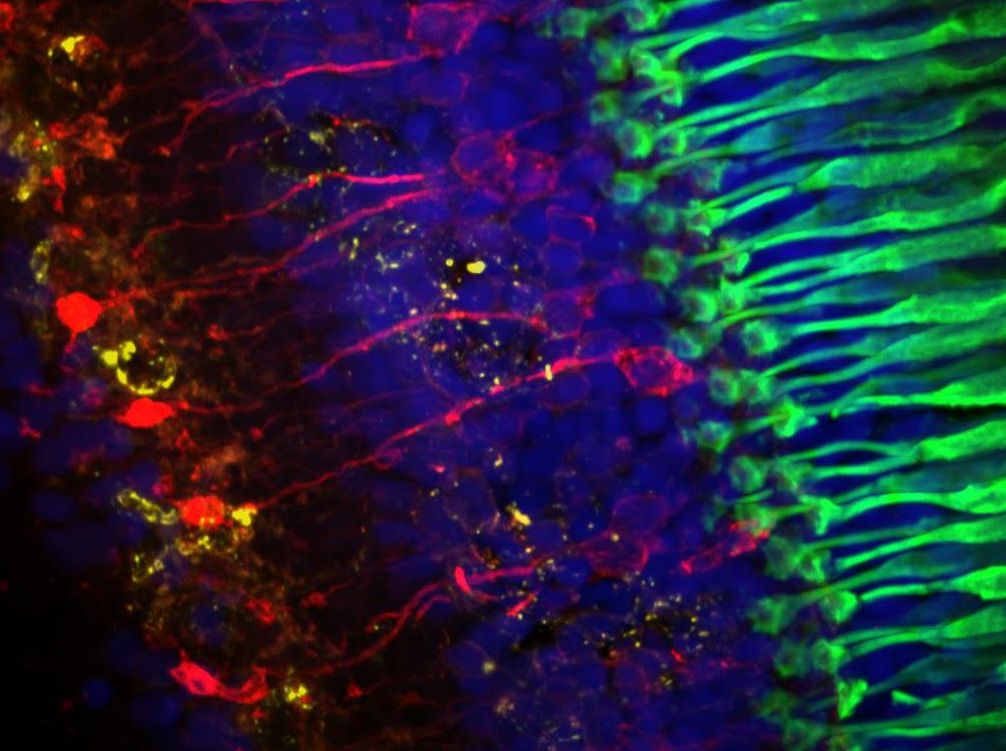
b - 3D reconstruction of tissue with MC(red, biocytin) and adult-born GC(green, ChR2-EYFP) cell nuclei (blue, DAPI). The white grid (8×8) shows the precise targeting of light application - the yellow outlined area is illuminated, the orange is not.
c - Postsynaptic voltage measurements corresponding to the illumination of a specific region (yellow/orange) from the 3D reconstruction(b) (Andor, 2016).
The Mosaic3 active illumination module has the following parameters:
- Illuminating multiple regions of interest simultaneously (DMD - digital mirror device).
- Incorporated memory for storing lighting patterns
- LEDlight source.
- Active lighting with a maximum speed of 5000 fps
- Minimum exposure time is 50-200 μs
- Compatible with most microscopes from the world's leading manufacturers
Report source: "The Plasticity behind Memory | Andor Mosaic | Active Illumination." Accessed December 7, 2016. www.andor.com


