Take a look at the new application with the data we took during the demonstrations of our system - the Andor Dragonfly confocal microscope. The multi-modal concept allows imaging of a wide range of samples, from miniature plant cell nuclei to entire root sections. It is also possible to image large sample volumes or multiple layers very quickly in time-lapse experiments.
Application: confocal microscopy of plant samples
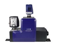
The configuration of the microscope was as follows:
- Confocal unit - Dragonfly 505
- sCMOS camera - Zyla 4.2 Plus USB 3.0
- EMCCD camera - iXon Ultra 888 SRRF Stream
- Acquisition software - Fusion (deconvolution/srrf stream/stitching)
- Analysis software - Imaris (3D visualization, video and image creation, analysis)
- Microscope body - Nikon Ti2 (inverted)
- Objectives - 10x, 20x MI, 40x Oil, 60x Oil , 100x Oil
- Excitation wavelengths - 405nm/488nm/561nm/637nm