The Andor Dragonfly confocal microscope is perfect for plant biologists. The multi-modal concept allows imaging of a wide range of samples, from miniature plant cell nuclei to entire root sections. It is also possible to image large sample volumes or multiple layers very quickly in time-lapse experiments.
Thanks to the zoom optics in front of the two cameras (1x, 1.5x, 2x), the effective pixel size can be adjusted according to the observer's requirements, thus achieving imaging below the Niquist criterion. The minimum effective pixel size can be e.g. 33 nm when using a Zyla camera with 2x zoom and 100x lens (1.49 NA) - calculations of the effective pixel size for the Dragonfly system - In very good quality, it is possible to penetrate deep into the sample, mainly due to the Borealis excitation beam guiding technology. Depending on sample preparation and quality, this can be up to hundreds of micrometers.
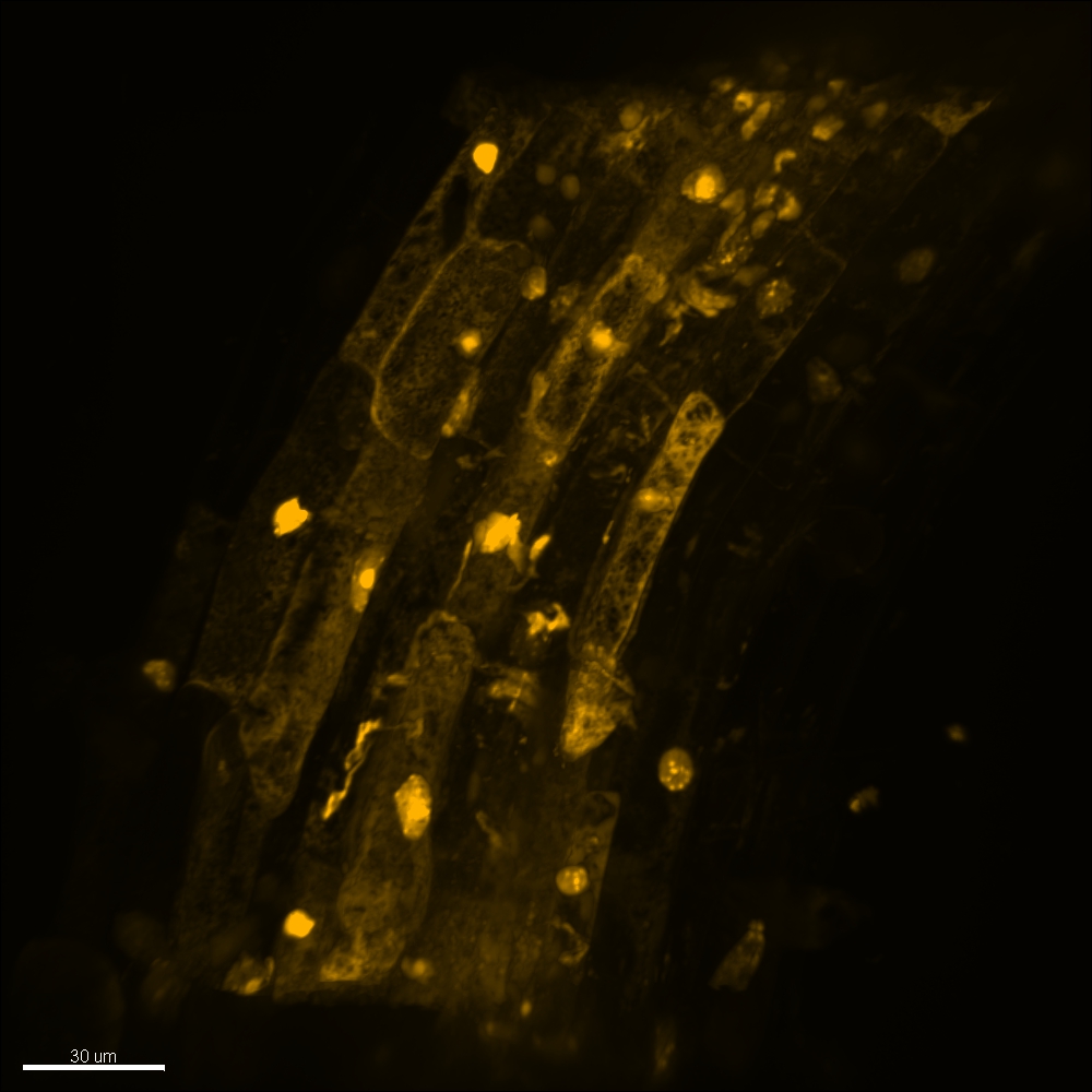
Figure 1: Root of a caterpillar (Arabidopsis) seedling stained with propidium iodide (PI) , Andor Dragonfly, Zyla, 60x, Oil, NA: 1.49, Voxel 0.100 * 0.100 * 0.170 um, X/Y: 2048*2048 pixels, Z: 389. Samples provided by Anna Nowicka from ÚEB, Olomouc, Czech Republic.
Since the Andor Dragonfly confocal microscope is very fast and flexible, several fields of view with high quality and penetration depth can be imaged in a short time. These fields of view are merged immediately after scanning by the FUSION software and the result can be, for example, a large part of a root imaged completely in very good resolution. Another advantage of Borealis technology is the high uniformity of illumination over a large field of view - this allows stitching without large overlaps and also quantitative measurement of the signal within the field of view.
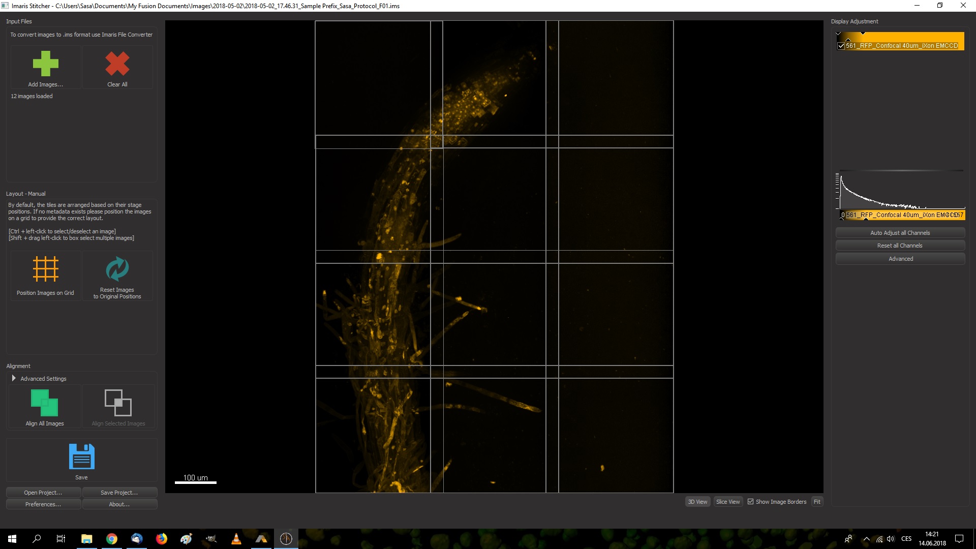
Figure 2: Root of a caterpillar (Arabidopsis) seedling stained with propidium iodide (PI) , Andor Dragonfly, iXon, 40x, Oil, NA: 1.3, Voxel 0.301 * 0.301 * 0.278 um, X/Y: 1024*1024 pixels, Z: 154. Imaris Stitcher - 12 full different fields before merging. Samples provided by Anna Nowicka from ÚEB, Olomouc, Czech Republic.
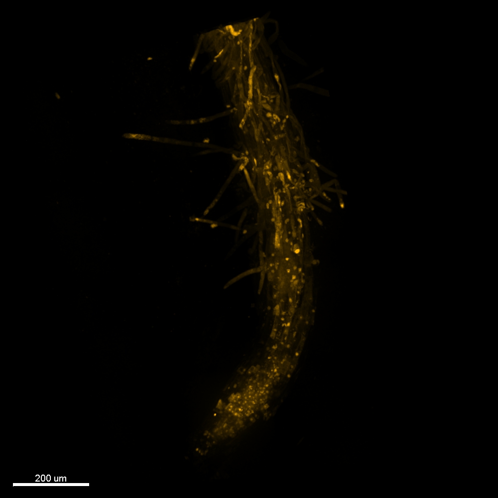
Figure 3: Root of a caterpillar seedling (Arabidopsis) stained with propidium iodide (PI) , Andor Dragonfly, iXon, 40x, Oil, NA: 1.3, Voxel 0.301 * 0.301 * 0.278 um, X/Y: 2879 * 3853 pixels, Z: 154. Connected using FUSION software immediately after scanning. Samples provided by Anna Nowicka from ÚEB, Olomouc, Czech Republic.
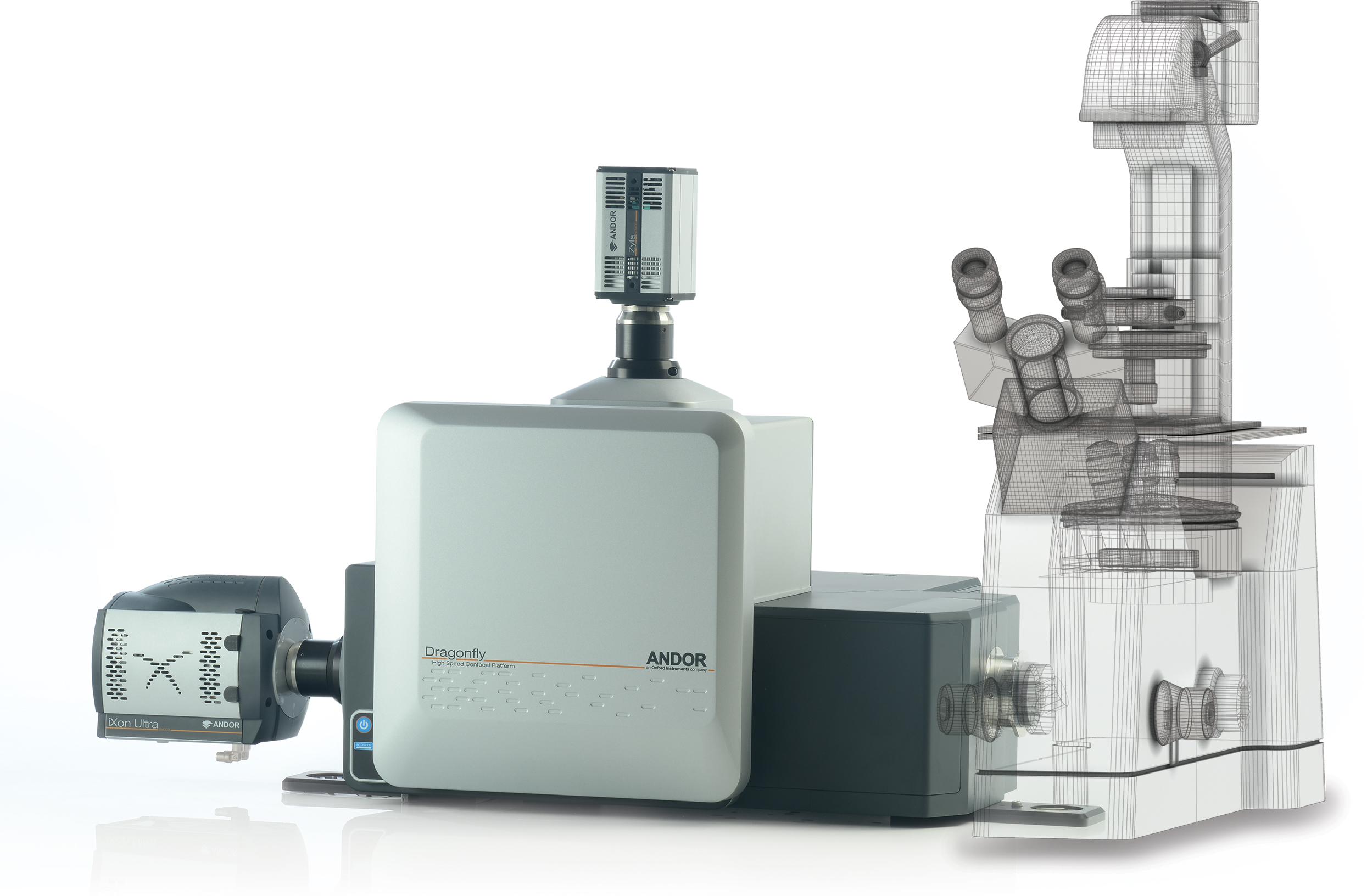
Confocal microscope Andor Dragonfly:
Scanning speed
- 400 frames per second (512 × 512 pixels)
- Minimum exposure - 2.5 ms
Sensitivity
- 90% quantum efficiency for iXon cameras (EMCCD)
- 82% quantum efficiency for Zyla cameras (sCMOS)
Microscope
- Precision XY motorized stage
- Very fast Z piezo in the range up to 500 um
- Hardware autofocus
- Dedicated lenses
Read more
- Borealis - greater efficiency and uniform sample illumination
- Stitching/montage - incorporated into Fusion acquisition software
- Active blanking - interfacing between light source and cameras