Confocal microscopy enables fast and high-quality imaging at high resolution. The acquired data in the form of optical sections must be reconstructed and subsequently analyzed. A3D colocalization study is a reliable method that reveals structural relationships between imaged objects.
Dr. Xavier Forns' laboratory (IDIBAPS&NIH) is researching the kinetics of the Hepatitis C virus to better understand the process of re-infection with this virus after liver transplantation. The main point of interest was to compare the expression of the Hepatitis C virus receptor with the overall kinetics of infection.
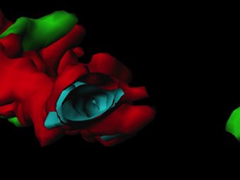
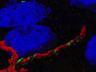
Figure 1: Liver cell membrane (3D visualization)
Imaris ) - colocalization of tight junction proteins, claudin - 1 (green) and occludin (red). Nuclei (blue).
Fixed biopsies of infected livers were imaged witha confocal microscope. Imaris software was used for 3D visualization and image analysis. The3Dcolocalization study focused on receptors and tightjunctions that may influence the mechanism of virus entry into the liver.
This study, together with other results, showed that the amount of Hepatitis C virus receptor at the time of liver transplantation regulates the initial kinetics of the virus. Hepatitis C virus reinfection after transplantation is directly linked to the amount of tight junctions in liver cell membranes.
Imaris software with the Coloc module offers a very intuitive and flexible setup for3D colocalization analysis:
- Manual or automatic adjustment of the sensitivity threshold(threshold).
- Intuitive 2D histogram with all voxel intensities
- Real-timestatistics (Pearson, Mander, % volume/ROI)
- Colocalization study of dynamic event images(timelapse)
- Creation of a new channel for data presentation
For more detailed information on the colocalization algorithms used in the Coloc module, please see the scientific paper - Costes et al. "Automatic and Quantitative Measurement of Protein-Protein Colocalization in Live Cells."
Webinars
"3-D colocalization study of viral kinetics." Accessed July 28, 2017. www.bitplane.com.


