The main group of proteins in the cell nucleus of eukaryotic cells are proteins that can bind to DNA in the form of chromatin (chromatin binding protein). It is the process of chromatin-binding proteins binding to DNA that is very dynamic and influences gene expression.
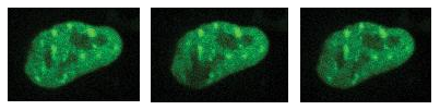
Figure 1: Mouse embryonic stem cell nucleus, green - H1 - YFP. Left - image of fluorescence signal from H1 protein. Middle - image showing the state after targeted illumination (photobleaching) by FRAP. Right - partial return of the fluorescence signal to the illuminated site (recovery).
Fluorescent tagging (fluorescent tag) using yellow fluorescent protein (YFP) was used to study the process of association and dissociation of chromatin-binding proteins (HP1, H1) on DNA in vivo. Fluorescence was observed in the nuclei of mouse embryonic stem cells by microscopy (Figure 1, left) confirming the presence of tagged proteins (HP1, H1). To study the dynamic processes taking place, a specific location in the observed nucleus - enabled by the active illumination module- was illuminated (photobleaching) using the FRAP method. Subsequently, thanks to the possibility of time-lapse imaging, a signal capturing chromatin remodeling and fluorescence signal change at the illuminated spot in the observed nuclei was obtained.
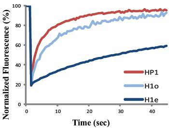
Figure 1: representation of FRAP targeting the HP1 and H1 proteins.
From the data obtained, the half-lives of association of HP1 and H1 proteins to chromatin were determined, as well as the differences in these half-lives for different forms of chormatin - heterochromatin/euchromatin
Possibilities of the active illumination module - Mosaic3:
- Illuminating multiple regions of interest simultaneously(DMD - digital mirror device)
- Incorporated memory for storing illumination patterns
- Light source: laser
- Active lighting with a maximum speed of 5000 fps
- Minimum exposure time is 50-200 μs
- Compatible with most microscopes from the world's leading manufacturers
"FRAPPA - Study the Dynamics of Chromatin Binding Proteins." Accessed December 5, 2016. www.andor.com.


